Leg Tendon Anatomy : Muscles Of The Leg And Foot Classic Human Anatomy In Motion The Artist S Guide To The Dynamics Of Figure Drawing : The hind leg can be confusing to some owners, but it has some of the same features as a human.
Leg Tendon Anatomy : Muscles Of The Leg And Foot Classic Human Anatomy In Motion The Artist S Guide To The Dynamics Of Figure Drawing : The hind leg can be confusing to some owners, but it has some of the same features as a human.. The main function of this muscle is to dorsiflex the foot, as well as invert it. This premium muscle model is life sized and comes on a sturdy base for easy display and study. See tendons muscles foot lower leg anatomy stock video clips. In the leg muscles diagram above, there are many muscles that make up your legs and support it to move. The gastrocnemius is the larger calf muscle, forming the bulge visible beneath the skin.
Most leg pain results from wear and tear, overuse, or injuries in joints or bones or in muscles, ligaments, tendons or other soft tissues. This muscle is part of the gluteal group which consists of three muscles. The muscular section closest to the body is the thigh, the middle section (the drumstick) corresponds to the human lower leg, and if the foot were still attached, it would follow the drumstick. These all work together to bear weight. Below are the muscle groups we will go over.
6 Muscles Of The Lower Leg Simplemed Learning Medicine Simplified from simplemed.co.uk Knee joint anatomy is complex with muscles, ligaments, cartilage and tendons. The quadriceps tendon works with the muscles in the front of your thigh to straighten your leg. Then we get to the hock; Below are the muscle groups we will go over. Anatomy of leg and foot human muscular. The achilles tendon is also located in the lower leg. Tendons consist of densely packed collagen fibers. The knee joint enables the movement of bending and straightening your legs.
The leg anatomy is so complex, containing both the knee and hip joints.
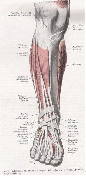
Then we get to the hock; See tendons muscles foot lower leg anatomy stock video clips. The knee joint is commonly injured, so understanding its anatomy can help you understand the conditions that cause problems, so you stay safe and prepared. Tendons are strong cords of fibrous tissue that attach muscles to bones. One of the most important tendons in terms of mobility of the leg is the achilles tendon. The tibialis anterior (tibialis anticus) is situated on the lateral side of the tibia; The origin of this nickname is obscure, but it may have to do with the practice of butchers of hanging the thighs of butchered animals such as pig (the hams) by the tendons of these three muscles. The hamstrings are three muscles at the back of the thigh that affect hip and knee movement. In the distal portion of the leg, the muscle fibers converge to form a tendon that extends through the medial side of the foot toward the muscle's insertion; This important tendon in the back of the calf and ankle stores the elastic energy needed for running, jumping, and other physical activity. Tendons consist of densely packed collagen fibers. Muscle anatomy gluteus 12 photos of the muscle anatomy gluteus gluteus muscle anatomy ct, gluteus muscle anatomy mri, human muscle anatomy gluteus maximus, muscle anatomy gluteus, muscle anatomy of gluteal, human muscles, gluteus muscle anatomy ct, gluteus muscle anatomy mri, human muscle anatomy gluteus maximus. Tendon and ligament injuries often go hand in hand with horses involved in vigorous athletic pursuits.
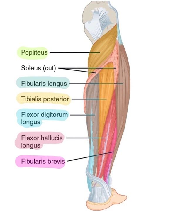
Horse hoof and leg anatomy: Hochwertiges kletterzubehör für dein outdoor abenteuer! Both cross the ankle, but the peroneus longus wraps underneath the cuboid crossing the plantar aspect of the foot as well, and inserts at the base of the first metatarsal. The technical term for a dog knee is the stifle joint. This muscle anatomy model shows full detail of the veins and arteries in the human leg.
Anatomy Of The Quadriceps Muscles from www.verywellfit.com Knee joint anatomy is complex with muscles, ligaments, cartilage and tendons. Most leg pain results from wear and tear, overuse, or injuries in joints or bones or in muscles, ligaments, tendons or other soft tissues. Tendons of the anterior compartment of the leg, the anterior tibial vessels, and the deep peroneal nerve pass under it. The major extensor tendon in the leg the carpal, pastern and coffin joints are extended by the lateral digital extensor. The tibialis anterior (tibialis anticus) is situated on the lateral side of the tibia; You've probably heard this mentioned more in horses. The hamstrings are three muscles at the back of the thigh that affect hip and knee movement. Below are the muscle groups we will go over.
It's also the largest joint in the body.
See tendons muscles foot lower leg anatomy stock video clips. The muscle is primarily responsible for dorsiflexion and inversion of the foot.12 The main function of this muscle is to dorsiflex the foot, as well as invert it. For a more detailed anatomy of the muscle, check out the following leg muscle diagrams posted below. The technical term for a dog knee is the stifle joint. This muscle anatomy model shows full detail of the veins and arteries in the human leg. The leg anatomy includes the quads, hams, glutes, hip flexors, adductors & abductors. Anatomy ankle anatomy ankle + ligament + tendon the foot anatomy human ankle anatomy 3d leg muscle lower leg anatomy leg articulation peroneal ankle muscles foot. The leg anatomy is so complex, containing both the knee and hip joints. Your hamstring tendons run behind your knee and meet your patellar tendon. This important tendon in the back of the calf and ankle stores the elastic energy needed for running, jumping, and other physical activity. Ebraheim's educational animated video describes the muscle and nerve anatomy of the lower leg.there are fourteen muscles within the lower leg. The quadriceps tendon works with the muscles in the front of your thigh to straighten your leg.
This important tendon in the back of the calf and ankle connects the plantaris, gastrocnemius, and soleus muscles to. Leg pain can also be caused by blood clots, varicose veins or poor circulation. The gastrocnemius is the larger calf muscle, forming the bulge visible beneath the skin. It's also the largest joint in the body. In the distal portion of the leg, the muscle fibers converge to form a tendon that extends through the medial side of the foot toward the muscle's insertion;
Muscles Of The Anterior Leg Myfootshop Com from www.myfootshop.com The hock is like the human ankle. Then we get to the hock; The medial cuneiform bone (medial and inferior surface), and base of the first metatarsal bone. The leg (crus) extends from the knee to the ankle and contains the tibia and f. The muscular section closest to the body is the thigh, the middle section (the drumstick) corresponds to the human lower leg, and if the foot were still attached, it would follow the drumstick. It is the junction of the thigh and the leg and is a hinge joint. The hip joint allows you to move and rotate your legs pelvic area in all directions. Hochwertiges kletterzubehör für dein outdoor abenteuer!
The calf muscle, on the back of the lower leg, is actually made up of two muscles:
The hip joint allows you to move and rotate your legs pelvic area in all directions. Learn how they work together to avoid injury and stay active. This premium muscle model is life sized and comes on a sturdy base for easy display and study. The achilles tendon is also located in the lower leg. Just as the pieces of a wing correspond to those in a human arm, the pieces of a chicken leg correspond to those in a human leg. Dog leg anatomy is complex, especially dog knees, which are found on the hind legs. The lower leg lies between the knee and the ankle. The knee joint is commonly injured, so understanding its anatomy can help you understand the conditions that cause problems, so you stay safe and prepared. The hind leg can be confusing to some owners, but it has some of the same features as a human. The leg (crus) extends from the knee to the ankle and contains the tibia and f. The tibialis anterior (tibialis anticus) is situated on the lateral side of the tibia; The hock is like the human ankle. The lower leg is a part of the lower extremity, or leg.
There are many muscles located in the lower leg, but there are three that are particularly well known—the gastrocnemius and the soleus, which are the most powerful muscles in the lower leg, and the anterior tibialis leg tendon. This muscle is part of the gluteal group which consists of three muscles.
/bodybuilder-working-leg-muscles-in-gym-1195621671-b74db540db4d47baab080f2ccf98d4a1.jpg)